
Case 1
Age: 22 years
Gender : Female
Clinical Detail :
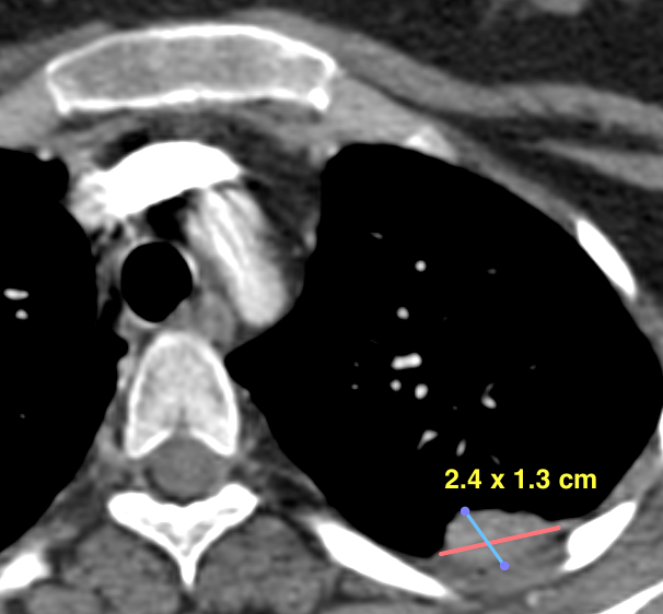
- Patient with osteosarcoma of right humerus with lung metastases.
- PET CT showed FDG avid posterior chest wall based LUL lung lesion measuring 2.4 x 1.3 cm. Lung function well preserved.
Procedure :
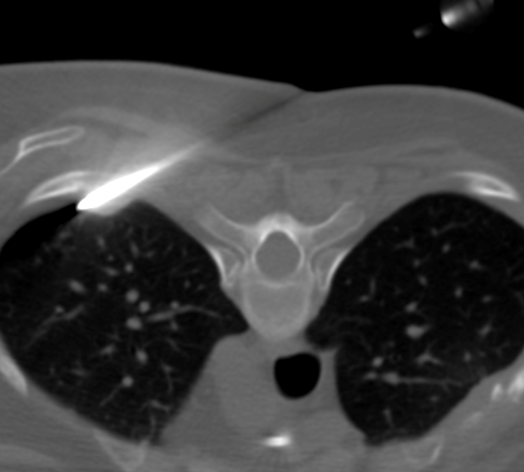
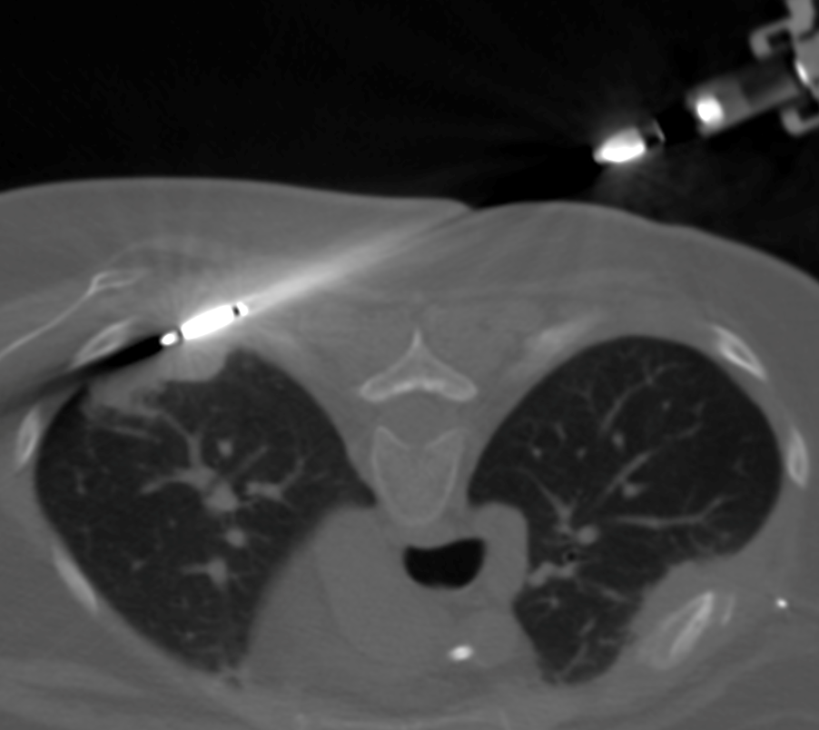
- Cryoablation done taking ablation zone of 28 x 29 mm with 2 x 10 minute cycles of freeze-thaw-freeze followed by 2.5 minutes of passive thaw.
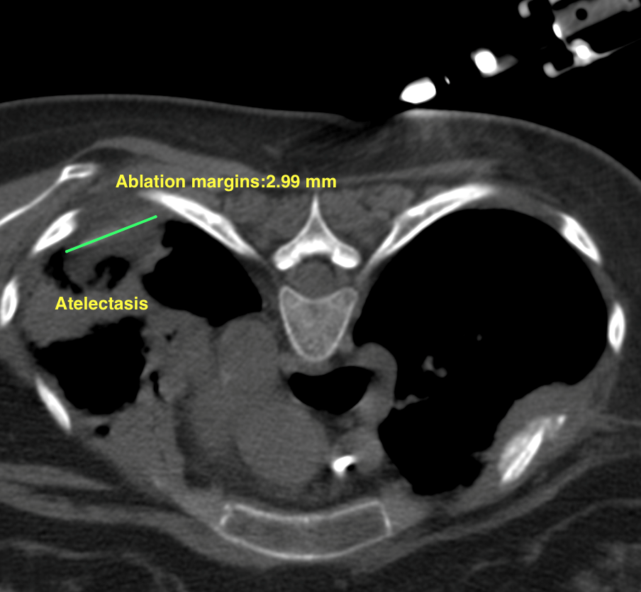
- Post-ablation check scan shows adequate ablation zone with surrounding atelectasis. No pneumothorax/hemothorax/chest wall hematoma seen.
Case 2
Age: 49 years
Gender : Male
Clinical Detail :
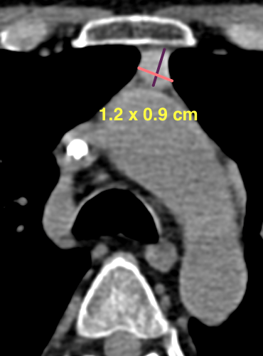
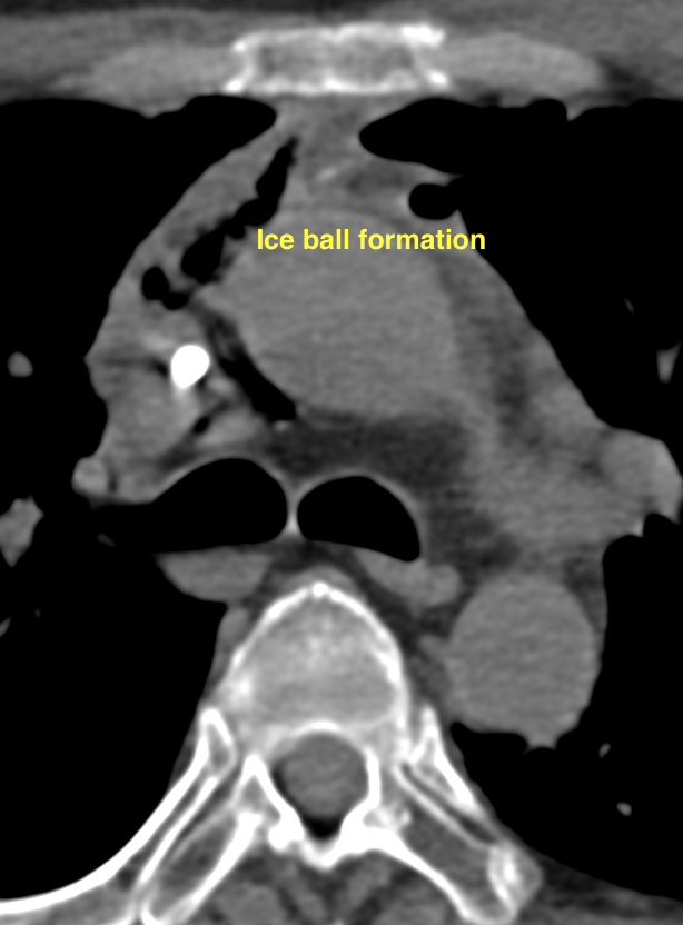
- Patient recurrent symptomatic thymoma.
- Previously treated with surgery.
- CT scan showed recurrent retrosternal nodule measuring 1.2 x 0.9 cm, close to aortic arch.
Procedure :
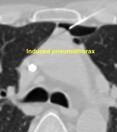
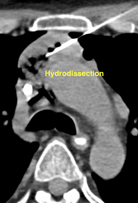
- Difficult access; induced pneumothorax and hydrodissection.
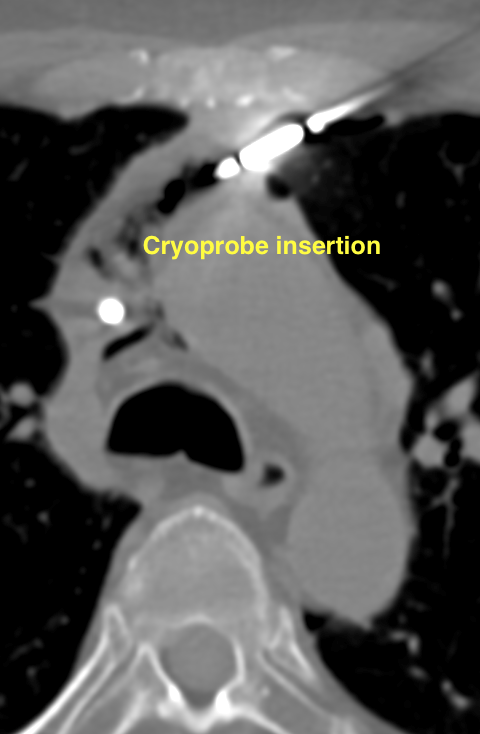
- 2 x 3 minute freeze-thaw freeze cycles with 13 G cryoprobe.
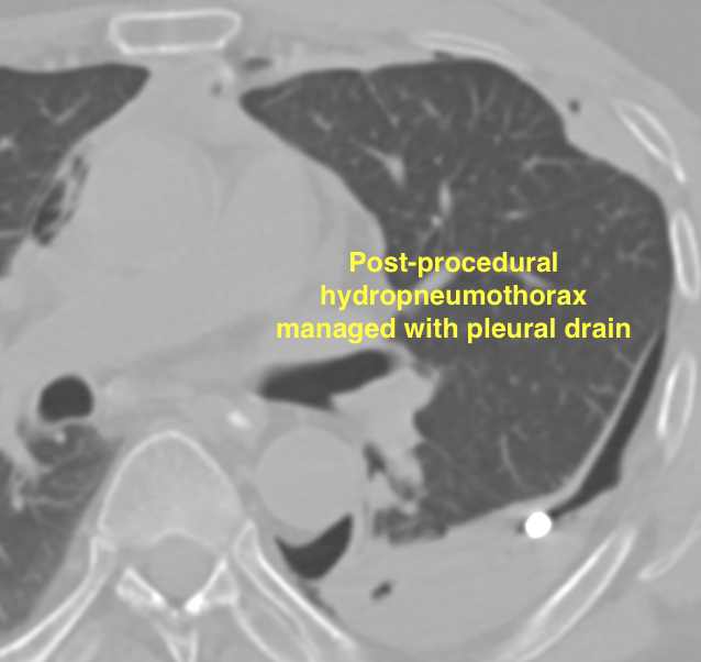
- Mild hydro-pneumothorax after procedure, inserted pleural drain which was removed next day. No injury to aortic arch.
Case 3
Age: 65 years
Gender : Female
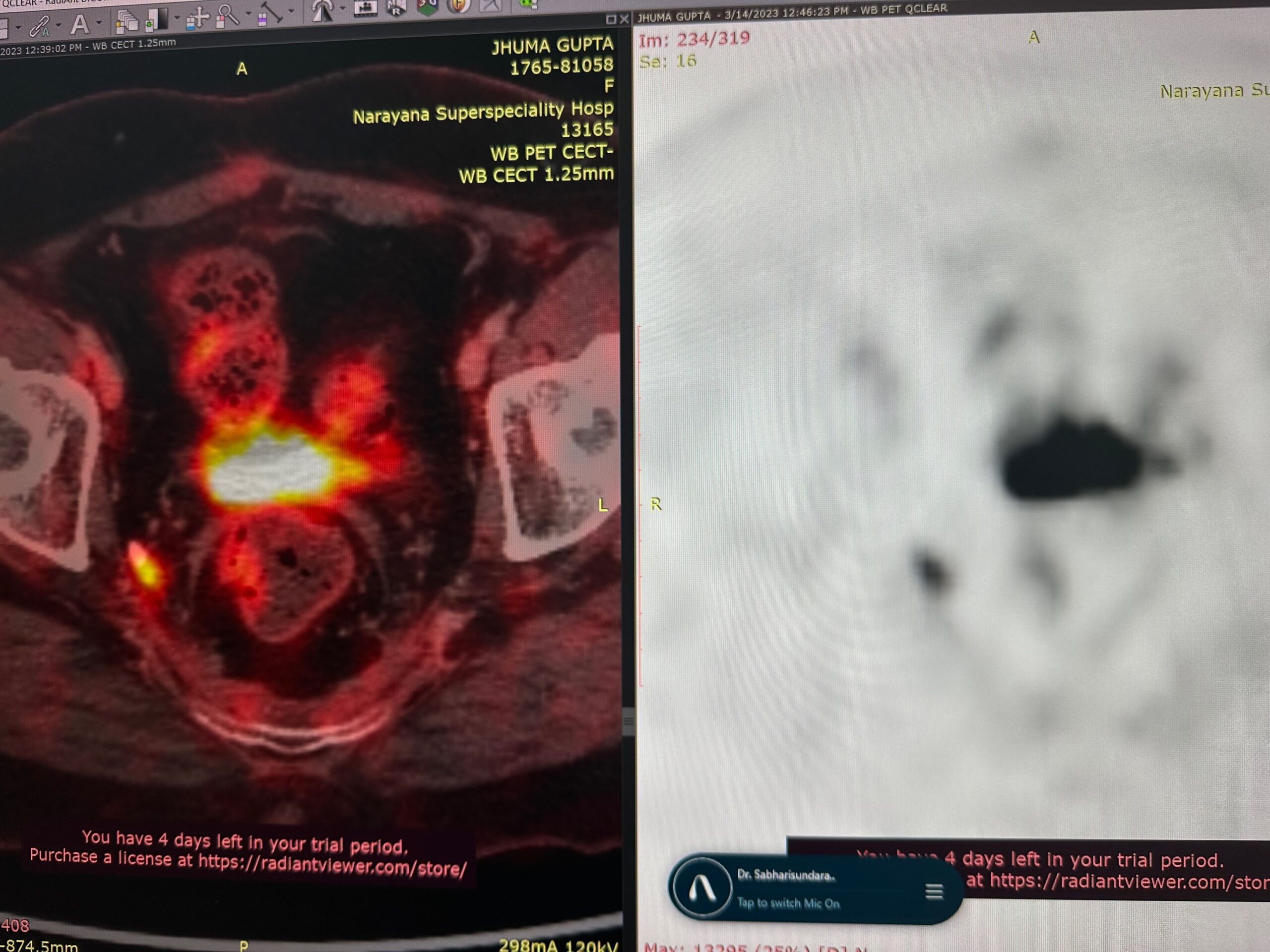
Clinical Detail :
- K/C/O adenocarcinoma of rectum post surgery + chemotherapy
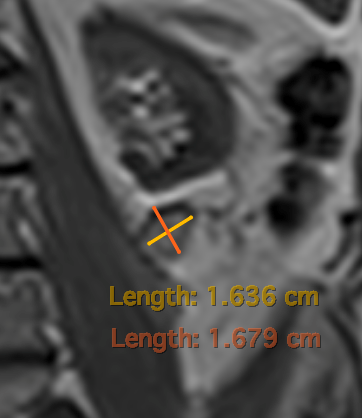
- Follow-up PET CT showed FDG avid nodule along right pelvic side wall
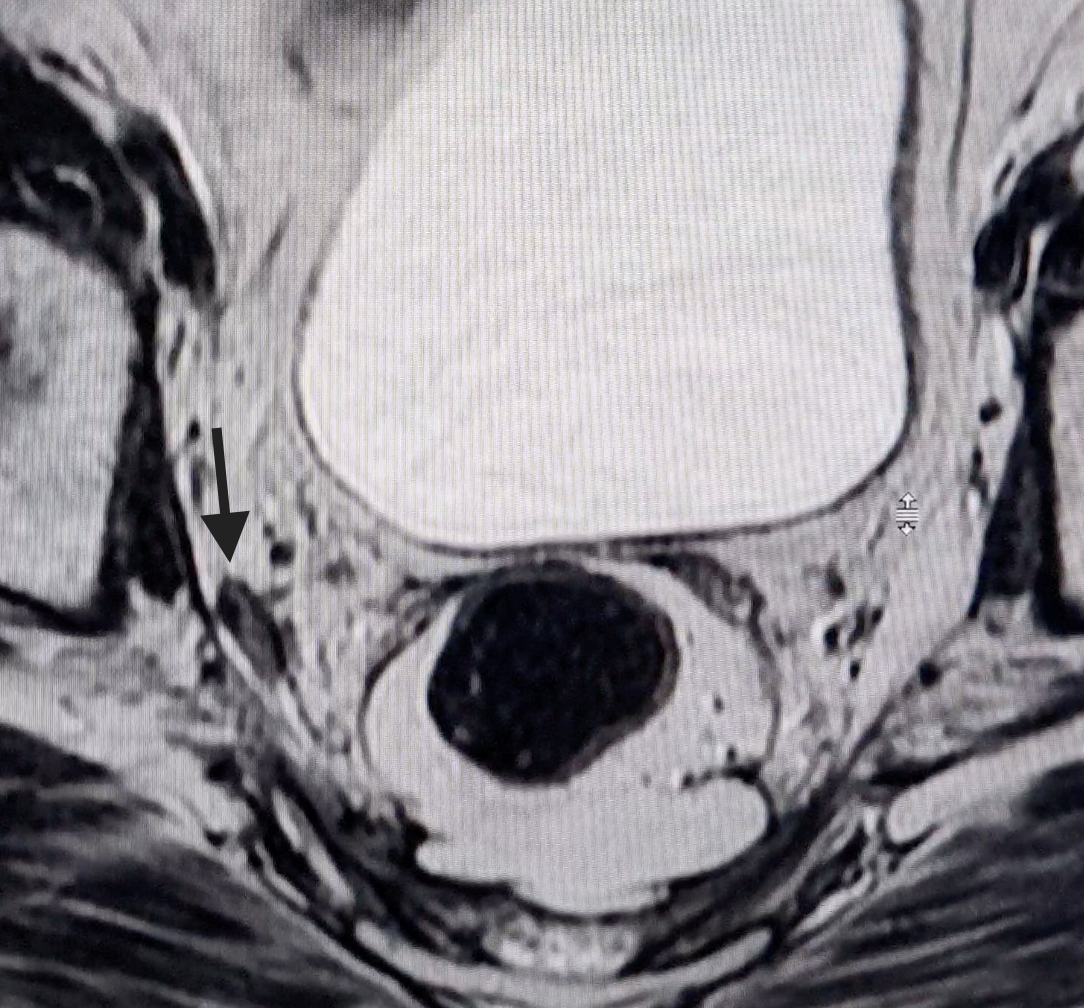
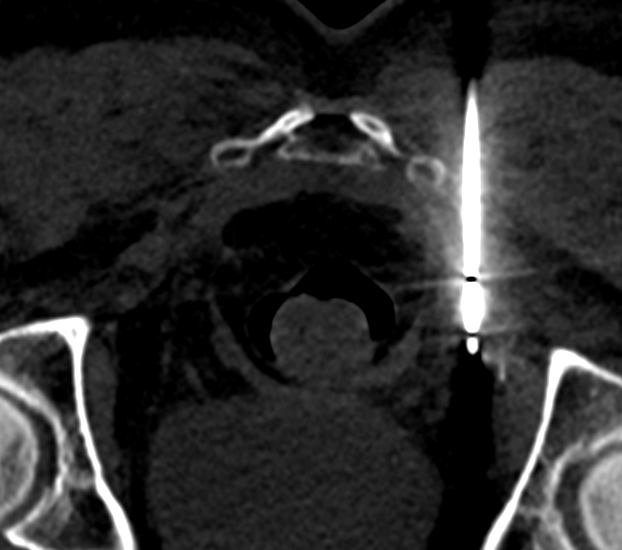
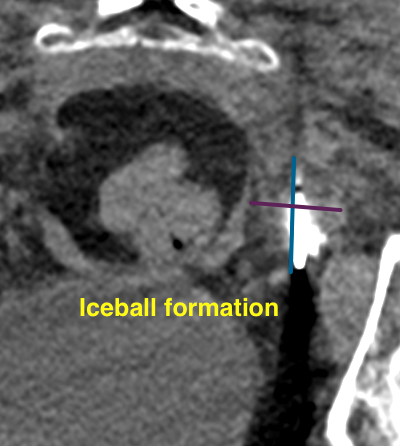
Procedures :
- 2 x 5 minute freeze-thaw freeze cycles with 13 G cryoprobe
- Adequate ablation margins with no post-procedure complications
Case 4
Age: 77 years
Gender : Male
Clinical Detail :
- K/C/O left renal RCC post partial nephrectomy 12 years ago. Otherwise asymptomatic
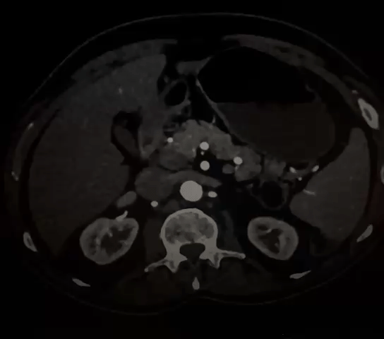
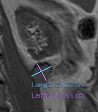
- On routine imaging, detected with enhancing nodule measuring 27 x 22 mm in resection bed close to left psoas
Procedure :
- Treated with cryoablation using 2 x 8 minutes freeze-thaw-freeze cycles.
- Adequate ablation margins with no post-procedure complications